Skeletal muscle constantly remodels itself to meet the changing demands of physical exertion, mechanical stress, and trauma. The muscle relies on a highly coordinated balance between cellular energy production and rapid repair mechanisms to maintain structural integrity and functional capacity. Disruption of this balance quickly leads to metabolic fatigue and tissue degradation.
Myocyte Enhancer Factor 2A (MEF2A) is a critical transcription factor that acts as the central manager for both metabolic efficiency and tissue repair.
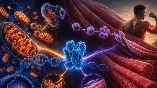
Metabolism: Keeps the mitochondria running efficiently so the muscle has the ATP (energy) it needs to contract and survive.
Regeneration: Activates muscle stem cells (satellite cells) to rebuild and replace damaged muscle fibers after a workout or an injury.
Usually, a cell treats these as two distinct jobs. MEF2A handles both. It turns on the genes required to generate cellular energy and the genes required to physically rebuild the muscle tissue.
Decoding the exact molecular role of MEF2A requires using an MEF2A ELISA kit to accurately track changes in MEF2A protein abundance in response to physiological stress and injury. MEF2A simultaneously governs mitochondrial quality control and orchestrates the activation timeline of muscle stem cells during regeneration.
MEF2A and Mitochondrial Homeostasis
Fueling the Muscle
Skeletal muscle is among the most metabolically demanding tissues in the human body. It requires a massive, uninterrupted supply of adenosine triphosphate (ATP) to sustain mechanical contraction.
To prevent cellular fatigue and metabolic collapse, high-performing muscles demand rigorous and constant mitochondrial homeostasis.
Mitochondria must be dynamically generated, maintained, and cleared in response to shifting cellular workloads. When metabolic stress increases, the cell relies on transcriptional networks to expand its energy capacity, a process heavily dictated by MEF2A.
Transcriptional Pathways
MEF2A drives mitochondrial biogenesis through a cooperative feed-forward loop with PGC-1α (peroxisome proliferator-activated receptor gamma coactivator 1-alpha).
PGC-1α is the master regulator of mitochondrial creation. MEF2A boosts the expression of this coactivator, fully known as peroxisome proliferator-activated receptor gamma coactivator 1-alpha. This spike sets off a cellular chain reaction. As a result, the cell begins transcribing the nuclear-encoded components needed to build the electron transport chain.
To keep up a steady supply of ATP during long workouts, the body relies on this pathway to drive oxidative phosphorylation.
A high-sensitivity MEF2A ELISA kit allows researchers to correlate protein abundance with downstream mitochondrial gene expression.
Quality Control & Mitophagy
MEF2A is also vital for removing aging or dysfunctional mitochondria, which is critical for mitochondrial quality control and mitophagy. When left unchecked, damaged mitochondria leak harmful reactive oxygen species (ROS), causing severe oxidative stress that damages muscle proteins and impairs contraction.
MEF2A helps maintain a healthy, efficient mitochondrial pool by regulating the clearance of damaged mitochondria that produce ROS.
The Molecular Orchestration of Muscle Regeneration
The Regeneration Challenge
Skeletal muscle regeneration relies heavily on a specialized population of muscle stem cells known as satellite cells. These cells reside in a dormant, quiescent state in healthy, resting tissue. However, these cells rapidly become active after mechanical trauma or severe injury.
Satellite cells rebuild damaged tissue by moving through three distinct phases:
- Activation: Awakening from their dormant state in response to injury.
- Proliferation: Rapidly dividing to multiply the cell population.
- Terminal Differentiation: Committing to become mature, functional muscle cells.
Synergy with MRFs
As these activated stem cells progress toward repairing the tissue, MEF2A emerges as a critical downstream coordinator. It works in close synergy with key Muscle Regulatory Factors (MRFs), such as MyoD and Myogenin, which are responsible for establishing muscle cell identity. It physically interacts with these master transcription factors, co-activating downstream target genes that force muscle progenitor cells to cease dividing.
They begin the process of differentiation, driving these individual cells to fuse together into elongated, multinucleated structures called myotubes. These myotubes ultimately mature into functional muscle fibers.
Temporal Dynamics
MEF2A expression is highly time-dependent. It does not remain consistently active. Its presence peaks precisely at the critical junction where progenitor cells transition from growing and proliferating to actively rebuilding structural tissue. Premature or delayed transcription factor activation can stall tissue repair. Researchers use an MEF2A ELISA kit to get the precise temporal mapping necessary to identify the exact therapeutic windows during myogenesis. Measuring these protein fluctuations allows researchers to track how efficiently the muscle is transitioning through the necessary phases of structural recovery.
Energy Demands Meet Structural Repair
The Metabolic Cost of Healing
Rebuilding damaged muscle tissue places a massive metabolic burden on cellular machinery. Repairing mechanical tears, synthesizing complex structural proteins, and facilitating the physically demanding fusion of progenitor cells into new myotubes requires an immediate and sustained surge of cellular energy. Without a massive supply of ATP, the entire regenerative cascade stalls. This vulnerability leaves the tissue susceptible to chronic weakness or incomplete repair.
The Dual-Action Role
The Structural System
It commands muscle stem cells to differentiate and fuse to physically rebuild the torn muscle tissue.
The Metabolic System
It activates mitochondrial biogenesis to generate the massive amount of ATP needed to fund that physical rebuilding.